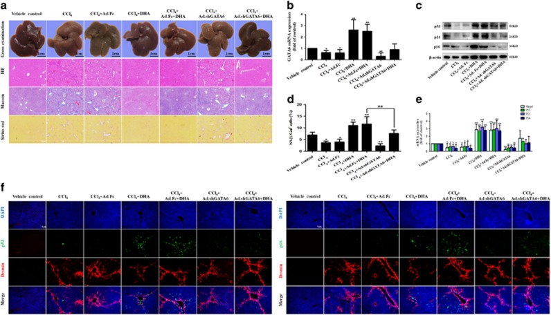
Figure 4.
DHA induces HSC senescence via a GATA6-dependent mechanism in vivo. Mice were randomly separated into seven groups: group 1, vehicle control (no CCl4, no treatment); group 2, model group (with CCl4, no treatment); group 3, Ad.Fc (a control adenovirus encoding IgG2 α Fc fragment) and CCl4-treated group; group 4, DHA (20 mg/kg) and CCl4-treated group; group 5, Ad.Fc, DHA and CCl4-treated group; group 6, Ad.shGATA6 (adenovirus encoding mouse GATA6 shRNA for inhibiting GATA6 expression) and CCl4-treated group; group 7, Ad.shGATA6, DHA and CCl4-treated group. (a) The pathological changes of the liver were observed by Gross examination, Scale bars are 1cm. Liver sections were stained with hematoxylin and eosin, Masson reagents and Sirius red. Representative photographs are shown. (b,c,e) Primary HSCs were isolated and subsequently were used to determine the expression of GATA6, p53, p21 and p16. (d) Liver sections were stained with β-galactosidase. (f) Liver sections were co-stained with desmin and p53 or p16. For the statistics of each panel in this figure, data are expressed as mean±S.D. (n=6); *P<0.05 versus control, **P<0.01 versus control, ***P< 0.001 versus control. ##P<0.01 versus CCl4+DHA+Ad.Fc treatment

