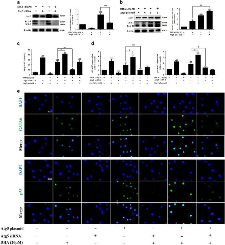
Figure 6.
Disruption of autophagy impairs DHA-induced GATA6 accumulation and HSC senescence in vitro. Activated HSCs were stably transfected with Atg5 siRNA or Atg5 plasmid, and then were treated with the indicated concentration of DHA for 24 h. (a, b) The transfection efficiency was confirmed by western blot analysis. (c) β-galactosidase staining analysis was used to detect HSC senescence. (d) Real-time PCR analyses were used to determine the expression of p53 and p16. (e) GATA6 and p53 immunostaining were used to determine the expression of GATA6 and p53. For the statistics of each panel in this figure, data are expressed as mean±S.D. (n=3); *P<0.05 versus control, **P<0.01 versus control, ***P<0.001 versus control. #P<0.05 versus DHA treatment, ##P<0.01 versus DHA treatment, ###P<0.001 versus DHA treatment

