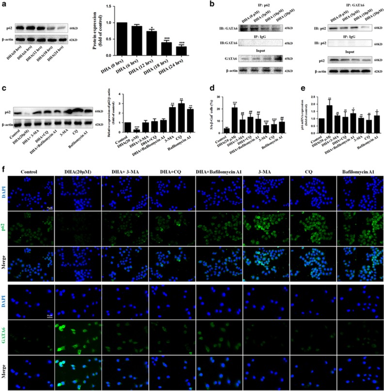
Figure 7.
Degradation of p62 is required for autophagy to mediate DHA-induced GATA6 accumulation and HSC senescence in vitro. (a) Activated HSCs were treated with DHA at 20 μM for various hours. Then, western blot was used to determine the expression of p62. (b) Immunoprecipitation assay was used to show the interaction of GATA6 and p62. (c) Activated HSCs were treated with 20 μM DHA in the absence or presence of autophagy inhibitors 3-MA (10 mM), CQ (10 μM), Bafilomycin A1 (5 nM). After 24 h incubation, the expression of p62 was determined by western blot analysis. (d) β-galactosidase staining analysis was used to detect HSC senescence. (e) Real-time PCR analyses were used to determine the expression of p16. (f) The expression of p62 and GATA6 was assessed by immunofluorescence. For the statistics of each panel in this figure, data are expressed as mean±S.D. (n=3); *P<0.05 versus control, **P<0.01 versus control, ***P<0.001 versus control. #P<0.05 versus DHA treatment, ##P<0.01 versus DHA treatment, ###P<0.001 versus DHA treatment

