Figure 1.
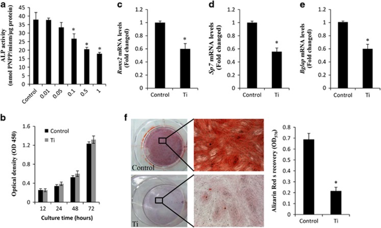
Ti particles inhibiting osteogenic differentiation. (a) MC3T3-E1 cells (9 × 10⁴ per well) were incubated in osteogenic medium containing various concentrations of Ti particles (0.01, 0.05, 0.1, 0.5 and 1 mg/ml) for 3 days. ALP activity was determined. (b) Cells (104/ml) were cultured with Ti particles (0.1 mg/ml) for 24, 48 or 72 h, and cell viability was determined by CCK-8 assay. mRNA levels of (c) runx2, (d) sp7 and (e) bgalp were determined using RT-PCR. (f) Matrix mineralization of differentiated MC3T3-E1 cells assessed by ARS staining. *P<0.05 versus control

