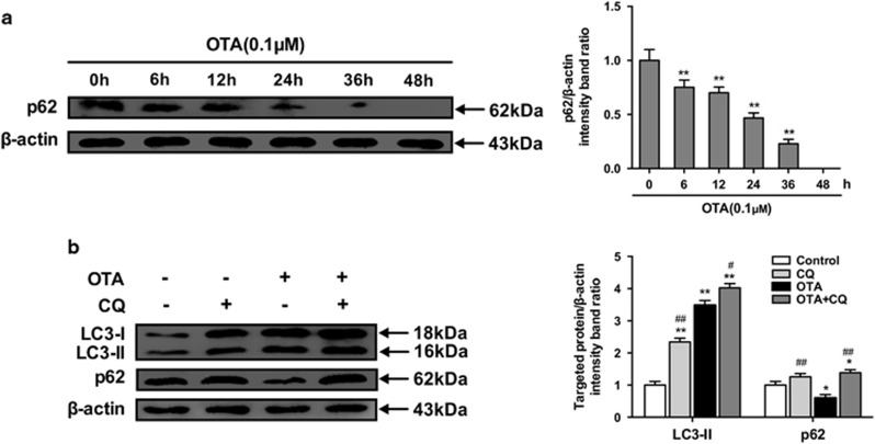
Figure 4.
OTA treatment enhances autophagic flux. (a) PK-15 cells were inoculated with PCV2 for 24 h and then inculated with OTA (0.1 μM). At the indicated times after inoculation, the cells were collected, and the expression of p62 and β-actin (loading control) was analyzed by immunoblotting with specific antibodies as described in Materials and Methods. The data are presented as means±S.E. of three independent experiments. Statistical significance compared with the control is indicated by *P<0.05 and **P<0.01. (b) PCV2-infected PK-15 cells were incubated with OTA (0.1 μM), CQ or with OTA and CQ for 48 h. After collecting the cells, the expression of LC3, p62 and β-actin (loading control) was analyzed by immunoblotting with specific antibodies as described in Materials and Methods. The data are presented as the mean±S.E. of three independent experiments. Statistical significance compared with the control is indicated by *P<0.05 and **P<0.01. Statistical significance compared with OTA is indicated by #P<0.05 and ##P<0.01

