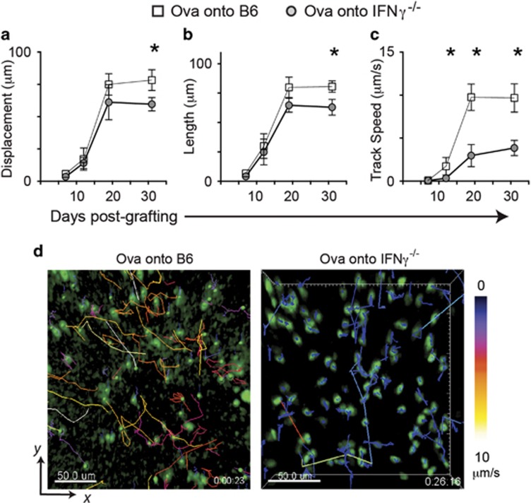
Figure 2.
Motility of CTL in tissues is enhanced by IFNγ. EGFP+CTL were adoptively transferred into B6 or IFNγ−/− mice before OVA skin was grafted. Post grafting, mice were anaesthetized and the graft edges imaged by 2-photon microscopy. Multiple sites per graft were imaged for 20-30 min each. EGFP+ cells were identified and tracked by software, and analyzed for CTL displacement (a), length of travel (b) and speed of travel (c) during the imaged time. Data are pooled from 4 regions imaged per graft, from 3 mice per time point. Averages are shown, error bars are SD. (*P<0.05) (d) Representative images of grafts on B6 recipients and IFNγ−/− recipients at day 19 post graft placement. CTL have been identified and tracked with track color coding for average speed of cell movement over the period of imaging. Bar, 50 μm (see also Supplementary Movies S1 and 2)

