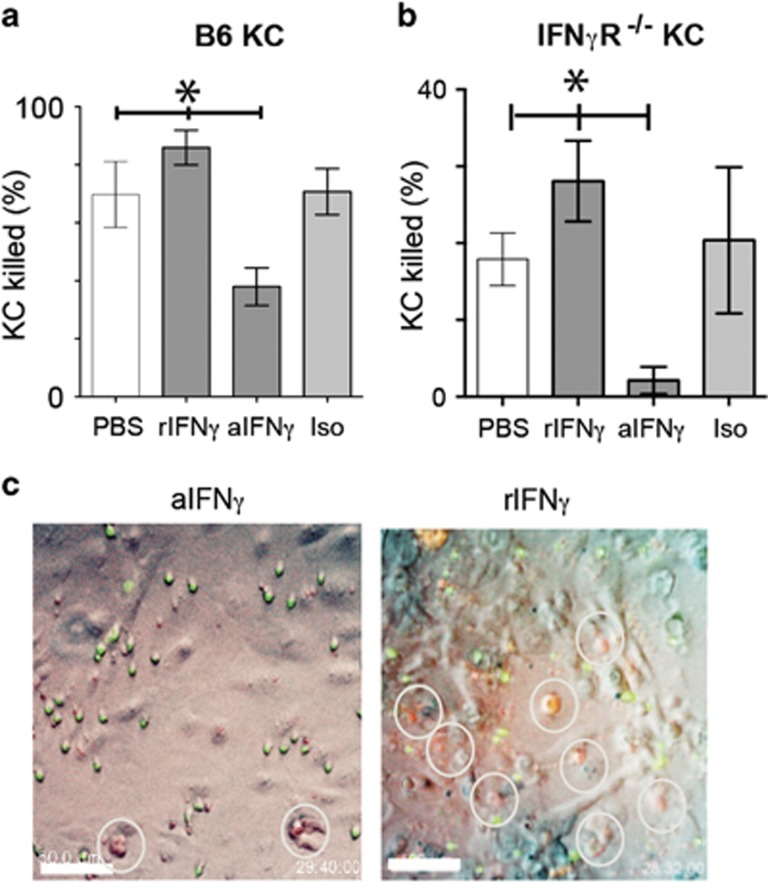
Figure 4.
Modulation of IFNγ signaling in CTL affects cytotoxic function. EGFP+OT-1 CTL were incubated with PBS, rIFNγ, anti-IFNγ or isotype antibody for 4 h, then washed thoroughly and added to SIINFEKL-loaded (a) B6 or (b) IFNγR−/− KC along with indicator dye for activated caspase. Co-cultures proceeded for 30 h, and were imaged by fluorescence time-lapse microscopy. Keratinocyte death was determined as in Figure 3. (*P<0.05, ‘NS’, not significant, ANOVA). (c) Frames of representative fields of co-cultures after 28 h of imaging. Merged red, green and brightfield images shown. Dead KC fluoresce red. EGFP+ CTL are green. Dead KC are shown by white rings. Scale bar is 50 μm

