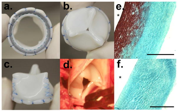
Figure 1.
Tissue-engineered aortic valve mounted on Sorin’s Mitroflow® Frame with a. bottom, b. top and c. side view shown, d. Top view of implanted valve in the ovine aortic positon, e. A trichrome cross section of the ovine fibroblast produced decellularized tissue and f. A trichrome cross-section of the human fibroblast produced decellularized tissue, with * representing the lumenal side, which becomes the ventricular side of the implanted valve. A 500 μm scale bar is shown in black on the trichrome sections.

