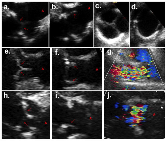
Figure 2.
Ultrasound imaging of the implanted aortic valve at time of implant with a. side view in closed position, b. side view in open position, c. end-on view in closed position, and d. end-on view in open position. At 12 weeks, side view in e. closed and f. open position, g. color Doppler side view showing unhindered flow through completely open leaflets. At 24 weeks, side view in h. closed and i. open position, j. color Doppler side view at 24 weeks showing unhindered flow through completely open leaflets.

