Figure 3.
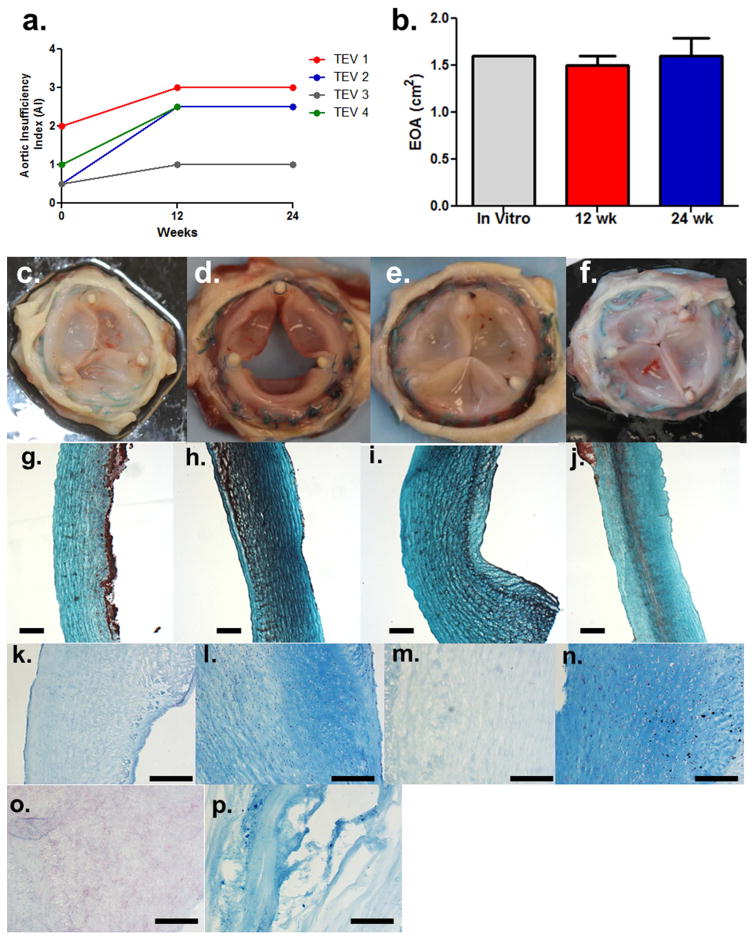
a. Aortic insufficiency grade over 24 week with 1/2/3/4 reporting trivial/mild/moderate/severe insufficiency, b. Measured EOA with average of all 4 valves at 12 week and 3 valves at 24 weeks. The 12 week values are based on replicate measurements of all valves (TEV1–4) and the 24 week values indicate the means and associated standard deviations based on 3 valves (TEV1,2,3). Top view of 24 week explanted valves c. TEV1, d. TEV2, e. TEV3, and f. 12 week explanted valve (TEV4). Trichrome-stained sections of the explanted valve leaflets for valves g. TEV1, h. TEV2, i. TEV3, and j. TEV4 showing organized collagen. Alcian blue staining showing k. proteoglycans in recellularized regions in leaflet of TEV2, but l. absent in regions still acellular, with similar staining pattern for m. recellularized and n. acellular regions in leaflet of TEV3, as well as o. the decellularized tissue pre-implantation. p. Positive control intervertebral disc for Alcian blue staining. Scalebars in all histology slides = 200 μm.

