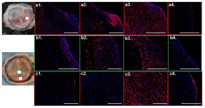
Figure 6.
Immunostaining of the explanted leaflets with top panel showing a1. CD45, a2. αSMA, a3. vimentin and a4. Von Willebrand factor staining for the 12 week explanted tissue (TEV4). The image of the valve on left shows the location from which the images are taken. The other two panels show images from the two regions of a 24 week explanted valve (TEV3) with image of the valve on left showing the locations, b1. and c1. CD45, b2. and c2. αSMA, b3. and c3. vimentin, b4. and c4. Von Willebrand factor. The white scalebar shown in all images = 200μm.

