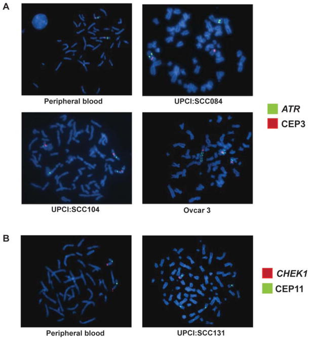
Figure 2.
(A) Two copies of the ATR gene (green) and two CEP3 (red) signals in a normal lymphocyte metaphase cell. (B) We observed a translocation of the ATR gene (green) in UPCI:SCC084. (C) In UPCI:SCC104, the ATR gene (green) is gained compared with CEP3 as a result of isodicentric chromosome 3 formation. (D) In the ovarian tumor cell line, OVCAR-3, we observe amplification of the ATR gene (green) compared with CEP3 (red). (E) A normal peripheral lymphocyte metaphase spread with two CHEK1 (red) and two CEP11 (green) signals. (F) UPCI:SCC131 OSCC cells with partial copy number loss of the CHEK1 gene (red) compared with CEP11 (green).

