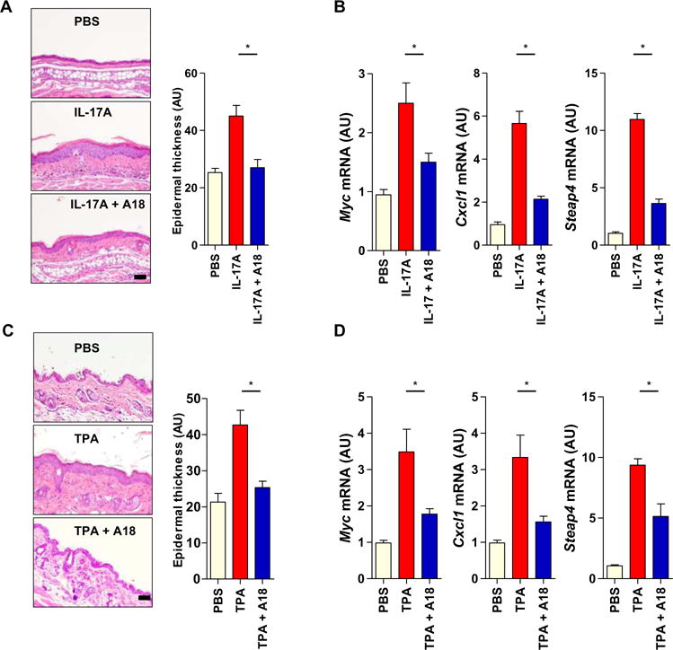
Fig. 3. A18 inhibits IL-17A–dependent skin hyperplasia in mice.

(A) Eight-week-old female C57BL/6J mice (n = 5 mice per group) were injected intradermally with PBS, IL-17A alone, or both IL-17A and A18. Left: Six days later, ear tissue was removed from the mice and subjected to hematoxylin and eosin (H&E) staining to assess ear thickness. Images are representative of three independent experiments. Right: Quantitative analysis of ear thickness in the indicated groups of mice. (B) Ear skin samples from the mice described in (A) were subjected to RT-PCR analysis of the relative abundances of Myc, Cxcl1, and Steap4 mRNAs. Error bars in (A) and (B) represent the SEM calculated from 15 mice from three independent experiments. (C) Eight-week-old female C57BL/6J mice (n = 6 mice per group) were treated on the dorsal skin with PBS, TPA alone, or both TPA and A18. TPA was applied to the mice on days 1 and 4, whereas A18 was applied daily. Left: On day 7, dorsal skin tissue was removed from the mice and subjected to H&E staining to assess epidermal thickness. Images are representative of three independent experiments. Right: Quantitative analysis of epidermal thickness in the indicated groups of mice. (D) Dorsal skin samples from the mice described in (C) were subjected to RT-PCR analysis of the relative abundances of Myc, Cxcl1, and Steap4 mRNAs. Error bars in (C) and (D) represent the SEM calculated from 18 mice from three independent experiments. *P < 0.05. Scale bars, 100 μm.
