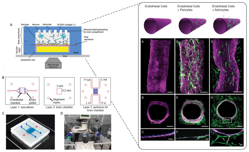
Figure 4.
Left panel A) Schematic representation of three compartments NVU. B) Microfluidic design layouts with details of each layer C) assembled device with top (blue) and bottom (red) channels. D) Experimental setup with microengineered NVU within environmental chamber for long term imaging. Reproduced from [62] with permission of AIP publishing. Right panel- Confocal imaging of various cell population within the cylindrical collagen lumen. Endothelial cells alone (A–C), with pre-added pericytes (D–F), or astrocytes in the bulk gel (G–I). Magenta is VE-Cadherin, green is F-actin, blue are nuclei. Arrows indicate contact points between cell populations. Reproduced from [63] with permission of PLOS.

