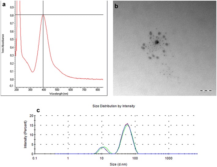
Fig. 1.

Characterization of silver nanoparticles. a UV–Vis analysis of silver nanoparticles showed peak absorption (0.806) at 395 nm, which matches with the surface plasmon resonance of silver nanoparticles, b TEM micrograph showing the morphology of silver nanoparticles: they are spherical with a mean size of 21 nm. (scale bar = 50 nm), c particle size distribution of the synthesized silver nanoparticles, showing two peaks: a smaller peak at 8.3 nm and a larger peak at 44.5 nm, which indicate presence of two particle size populations
