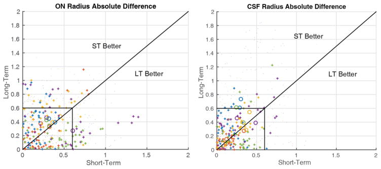
Figure 4.
Comparison of short- and long-term scan-rescan absolute error for the ON (left) and CSF (right). Large circles indicate the mean absolute error for a given nerve. Dots indicate individual points between nerves with the color corresponding to each subject. Pluses are individual points between nerves within the central third of the length of the nerve, the area which is most accurately imaged. The lines are drawn along unity and at resolution (0.6mm). Note that pluses tend to be localized within the box indicating reproducibility within a voxel for the central third of the nerve.

