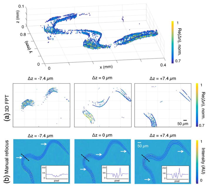
Fig. 6.
Tomographic reconstruction of a Trichinella spiralis parasite. (a) The worm’s curved trajectory resolved within various z planes. (b) Refocusing the same distance to each respective plane does not clearly distinguish each in-focus worm segment (marked by white arrows). Since the worm is primarily transparent, in-focus worm sections exhibit minimal intensity contrast, presenting significant challenges for segmentation (see intensity along each black dash in inset plots, where black dash location is in-focus in left image). FPT, on the other hand, exhibits maximum contrast at each worm voxel. See Visualization 1.

