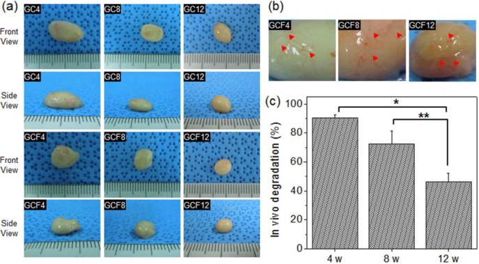
Figure 5.
(a) Images of the removed MCL without (GC) or with (GCF) osteogenic factors, after 4, 8, and 12 weeks. The numbers indicate the implantation time; (b) enlarged images of the removed MCL hydrogels (GCF) and (c) volume change in the in vivo excised MCL hydrogels (GCF) after 4, 8, and 12 weeks (*p < 0.001, **p < 0.05).

