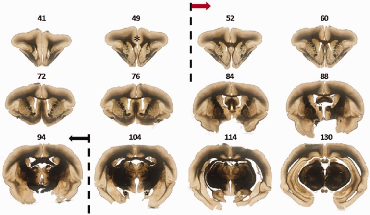
Figure 2.
The representative serial sections showing the range of ACC in tree shrew. Sample sections from a stack of 100-µm serial coronal sections. In No. 49 section, prefrontal cortex was indicated by asterisk. Sections from No. 52 to 94 showed the forebrain containing ACC, in which the ACC were framed by dash lines. Sections from No. 104 to 130 showed the forebrain containing posterior cingulate cortex, in which the hippocampus were indicated by stars.

