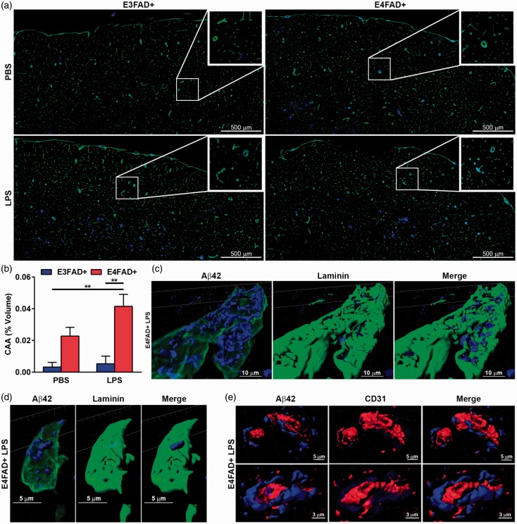
Figure 6.
CAA-like deposition is higher in E4FAD+ mice. (a) Mosaic images of 12 µm sections costained for laminin (green) and Aβ (blue) stitched together from individual 20× images. (b) Quantification of Aβ-laminin costaining. (c to e) 3D reconstruction from 63× confocal image of a cortical (c) larger vessels or (d, e) capillary stained for Aβ (blue) and (c, d) laminin (green) or (e) CD31 (red) indicating that CAA-like deposition is outside of the blood vessel within the basement membrane. (b) *p < .05 by two-Way ANOVA followed by Bonferroni post hoc comparisons comparing all groups.

