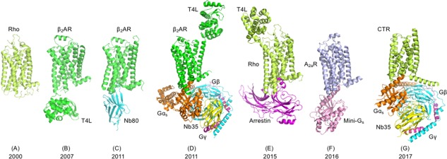
Figure 1.

Milestones of GPCR structure determination: PDB codes are given in parentheses. (A) Rhodopsin (1F88), (B) β2‐adrenergic receptor (2RH1), (C) β2AR with nanobody (3P0G), (D) β2AR with heterotrimeric G protein (3SN6), (E) Rhodopsin with arrestin (4ZWJ), (F) Adenosine A2a receptor with mini G protein (5G53), (G) Calcitonin receptor (CTR) with heterotrimeric G protein (5UZ7). Structures shown in panels A–F were solved by X‐ray crystallography, whereas the structure shown in panel G was solved by single particle cryo‐EM. The T4 lysozyme fusion partner facilitating crystal formation is indicated as T4L. Individual protein chains of receptor complexes are labeled and shown in different colors. Publication years are given at bottom of figure.
