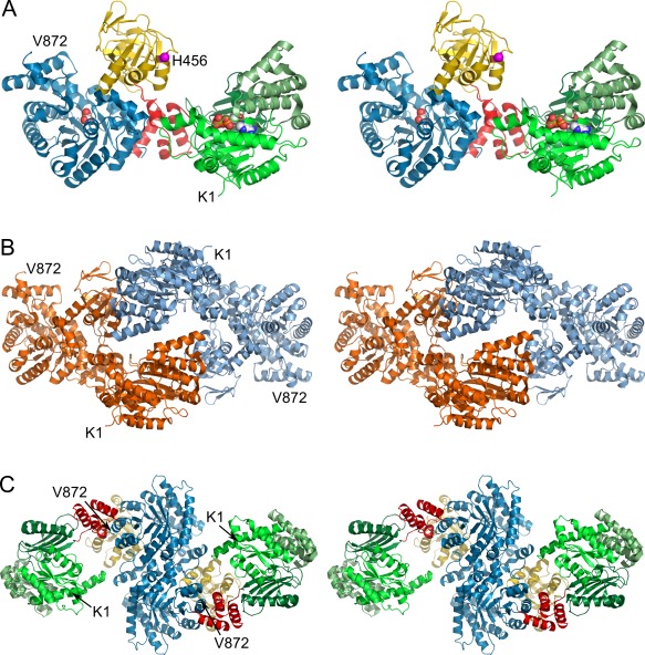
Figure 1.

(A) Stereo cartoon representation of 5LU4 chain A illustrating the overall domain organization. The nucleotide binding domain (NBD, aa 1–340) and its three subdomains are colored in different greens. The PEP/pyruvate binding domain (PBD) is colored in blue (aa 534–872). The central domain (CD, yellow, aa 381–516) with the catalytic His456 (magenta, shown as sphere) is attached to both substrate binding domains via two short linker helices (red, aa 341–380 and 517–533). Pyruvate and ADP bound to the PBD and NBD respectively are depicted as spheres. (B) Dimeric assembly within the asymmetric unit (ASU). The dimer is formed by contacts between the NBDs and CDs of chains A and B, colored orange and blue respectively. (C) Biological assembly as identified by the program EPPIC 11 and reconstructed from crystal symmetry. The dimerization interface is formed by both PBDs as previously described 12. Individual domains are colored according to (A). An interactive view is available in the electronic version of the article
