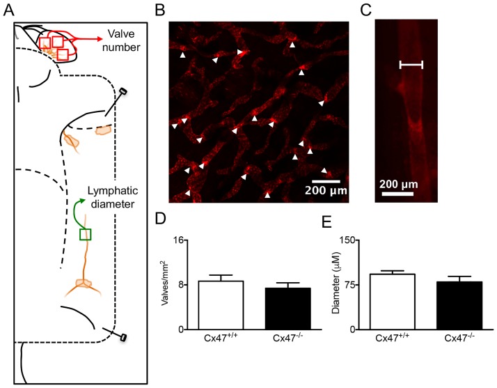
Fig 2. Normal lymphatic vessel organization in Cx47-deficient mice.
A) Schematic illustration showing localization of mOrange2 positive lymphatic vessels in a Cx47+/+Prox1tg/+ mouse. As indicated (red boxes), the number of lymphatic valves was quantified in three separate images covering > 75% of the inner ear surface. Moreover, lymphatic diameter was quantified in the lymphatic vessel draining the inguinal lymph node (green box). B) Representative image showing lymphatic valves (arrowheads) in the inner ear of a Cx47+/+Prox1tg/+ mouse. C) Representative image showing the measurement of the lymphatic diameter in a Cx47+/+Prox1tg/+ mouse at equidistance between two valves. D) Quantification of the number of lymphatic valves in Cx47+/+Prox1tg/+ (white bar) and Cx47-/-Prox1tg/+ (black bar) mice. E) Quantification of the lymphatic diameter in Cx47+/+Prox1tg/+ (white bar) and Cx47-/-Prox1tg/+ (black bar) mice. N = 7.

