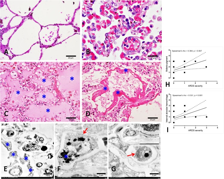
Fig 1. Histopathological and ultrastructural appearance in normal and severe malarial lungs.
A: normal lung represented by a very thin alveolar septum and clearly defined alveolar sac in contrast to the non-PE lung (B) which was characterized by thickened alveolar septum congested with blood components (e.g. PRBCs and WBCs). Apart from the thickened membrane, fluid (C*) and the hyaline membrane (D*) were deposited in the PE and ARDS lungs, respectively. Fine morphology of hyaline membrane (E*), macrophage (F; arrow)-ladened hemozoin pigment (F*) and PRBCs (G; arrow) with endothelial cell damage (G-inset) were frequently observed in the ARDS lung patients. A positive correlation between some histopathological changes and ARDS severity was calculated using Spearman test (H-I).

