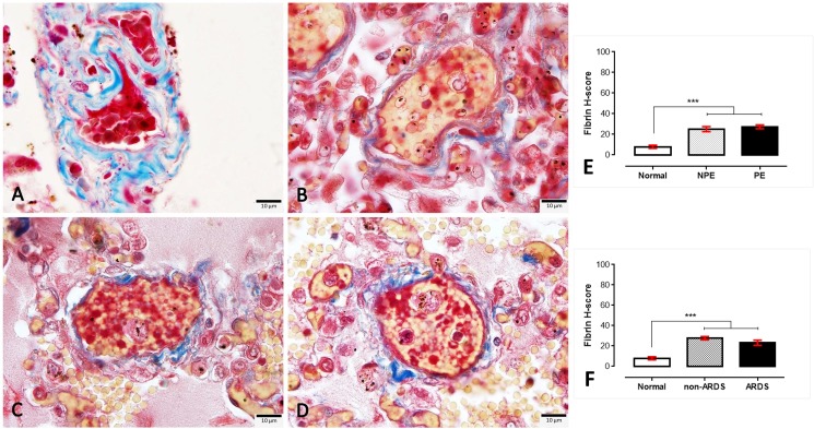
Fig 6.
Masson’s trichrome staining for intravascular fibrin deposition in normal (A), non-PE (B), PE (C) and ARDS (D) lungs. The fibrin staining is exhibited in the yellowish material found in the small blood vessels, whereas red blood cells and collagen fibrils were stained red and blue, respectively. The H-score expression is demonstrated by bar graphs for PE (E) or ARDS (F) lungs, to compare those expression Friedman test was performed (***; p-value <0.0001).

