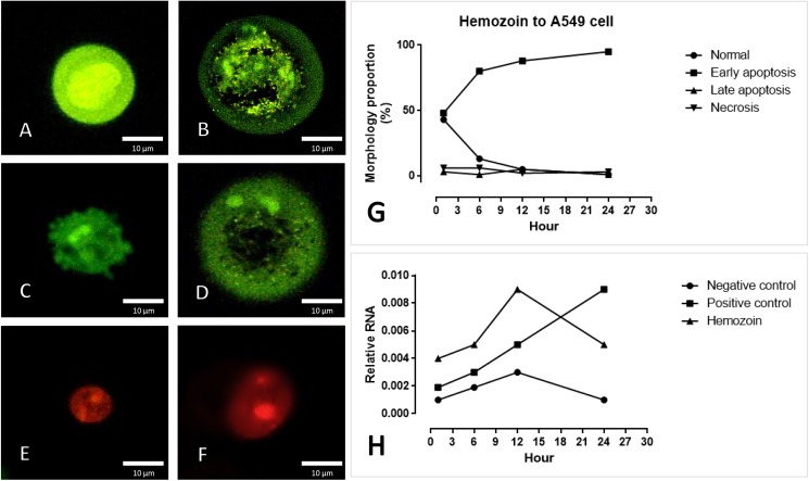
Fig 9. Apoptosis indicated by EB/AO staining and real-time RT-PCR.
S-hemozoin-induced pneumocytic apoptosis was characterized by morphological changes and staining patterns that included normal cells (A), early apoptotic cells with nuclear fragmentation or condense chromatin (B), membrane blebbing (C), cytoplasmic vacuolization (D), late apoptotic cells with condensed chromatin (E) and necrotic cells (F). The bar graph demonstrates that early apoptosis was predominately found following hemozoin treatment between 1–24 h with an increasing trend (G) in accordance with the mRNA expression of CARD-9 (H).

