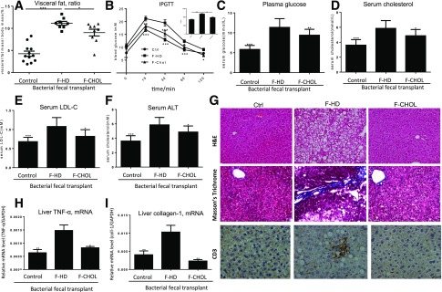
Figure 4.
FBT demonstrates that the microbes from the cholestyramine treatment are sufficient to antagonize the metabolic disorders of the recipient mice. The mice fed with HD or the control chow for 20 weeks were used as two types of recipients, which were subjected to gavage transplant from the donor microbes derived from cholestyramine-treated (F-CHOL) or HD (F-HD) mice (n = 10). A: Percent visceral fat deposition among the body mass, of the mice at the end of FBT treatment. B: IPGTT and area under the curve (AUC) of the mice at the end of FBT treatment. C: Fasting blood glucose of the mice at the end of FBT treatment. D: Plasma cholesterol. E: Plasma LDL-C. F: Serum ALT levels for liver injury. G: Hematoxylin-eosin (H&E) staining of liver tissues for hepatic steatosis; Masson’s trichrome staining of liver tissues for fibrosis; immunohistological staining of CD3+ lymphocytes for liver inflammation of the mice at the end of FBT. H: Liver inflammation was assessed by expression of TNF-α by the mice at the end of FBT. I: Liver fibrosis was assessed by expression of type I collagen. * or #, P < 0.05; ** or ##, P < 0.01. Comparisons were made against the F-HD group. Data were mean ± SEM.

