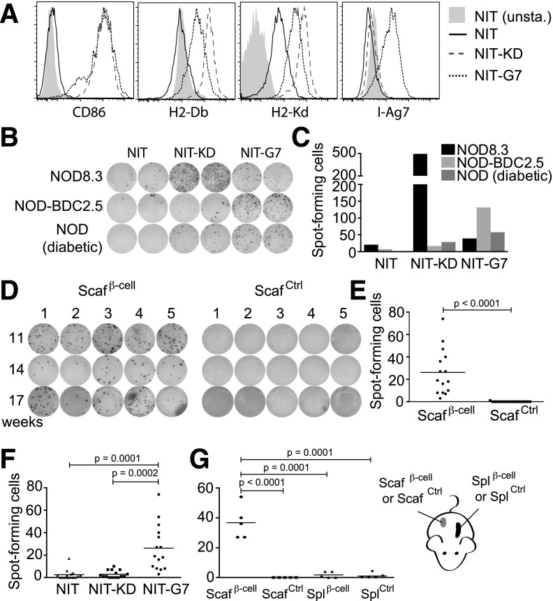
Figure 4.
β-Cell scaffolds enrich T cells specific for β-cell proteins. A: NIT-1 cells (NIT) were transduced with CD86 and either NLRC5 (NIT-KD) or CIITA (NIT-G7) and were analyzed by flow cytometry for expression of CD86, MHC class I (H2-Db and H2-Kd), and MHC class II (I-Ag7) or left unstained (NIT unsta.). B: Splenocytes (5 × 10⁵/well) from NOD8.3, NOD-BDC2.5, and diabetic NOD mice were analyzed for IL-2 secretion in an ELISPOT assay in response to live NIT-1, NIT-KD, and NIT-G7 cells (1 × 10⁵/well). T cells from NOD-BDC2.5 and NOD8.3 transgenic mice express TCRs that recognize peptides derived from the β-cell proteins on I-Ag7 and H2-Kd, respectively. C: Quantification of IL-2+ T-cell spots in B by using an ELISPOT analyzer. D: β-Cell and control scaffolds were removed from the backs of 11-, 14-, and 17-week-old NOD mice at 14 days postimplantation. Cells were extracted from the scaffolds and tested for IL-2 production in response to live NIT-G7 cells in an ELISPOT assay. E: IL-2 production was quantified with an ELISPOT analyzer. F: IL-2 production measured in response to live NIT, NIT-KD, and NIT-G7 cells from three cohorts of mice (11, 14, and 17 weeks old). G: T cells (7 × 10³/well) from β-cell scaffolds, control scaffolds, or spleens (Spl) from mice implanted with either β-cell scaffolds or control scaffolds were assayed in an IL-2 ELISPOT on live NIT-G7 cells and quantified. Scafβ-cell, β-cell scaffolds; ScafCtrl, control scaffolds.

