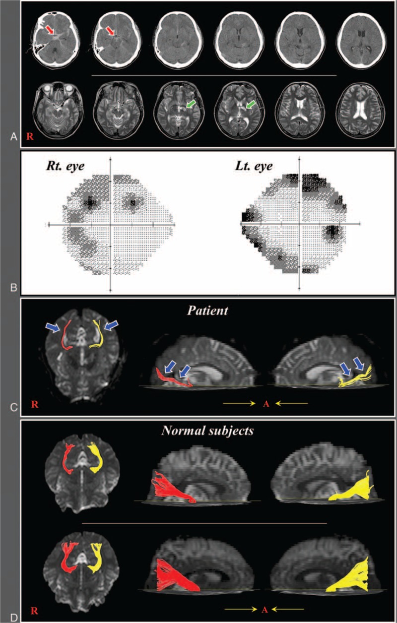
Figure 1.

(A) Brain CT images at onset show subarachnoid hemorrhage and T2-weighted MR images at 4 weeks after onset show a leukomalactic lesion in the left internal capsule. (B) The Humphrey visual field test of the patients shows a peripheral field defect in both eyes. (C) Diffusion tensor tractography images of the optic radiation in the patient (red color: right optic radiation, yellow color: left optic radiation). (D) Diffusion tensor tractography images of the optic radiation in 2 control subjects (62-year and 60-year old females).CT = computed tomography, MR = magnetic resonance.
