Fig. 1.
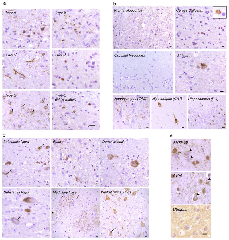
Histopathologic characteristics of FTLD-TDP type E. a Representative images of p409/p410 immunostained neocortical sections (layer II) from type A, B, C, D and E are shown (scale bar 20 μm). Type A shows dense neuronal cytoplasmic inclusions including ring inclusions, and neurites affecting superficial neocortical layers. Type B shows compact neuronal cytoplasmic inclusions with few neurites and involves superficial and deep neocortical layers. Type C shows long dystrophic neurites. Type D is typified by numerous intranuclear inclusions. Type E exhibits GFNI’s and grains in superficial and deep neocortical layers. Curvilinear oligodendroglial inclusions are also present (white matter). b Type E pathology is seen throughout most of the cerebrum including frontal neocortex, major white matter tracts such as the corpus callosum (inset shows high power image of curvilinear oligodendroglial inclusion), deep grey structures such as the striatum, and limbic regions such as the hippocampus (DG = dentate gyrus). Also shown is the occipital neocortex which is relatively spared (scale bars 10 μm). c Type E pathology is seen throughout the brainstem and cervical spinal cord. Shown are representative images from the substantia nigra, basis pontis (pons), medullary olive, dorsal medulla and ventral spinal cord. More compact or dense inclusions highlighted with arrows. Oligodendroglial inclusions highlighted with asterisks. Scale bars 10 μm. d Representative images of type E cases stained with anti-TDP-43 antibodies 6H6E12 and 5104, and the anti-ubiquitin antibody MAB1510 are shown (scale bar 10 μm). TDP-43 antibodies reveal both GFNI’s and grains with nuclear clearance of normal nuclear TDP-43 in affected neurons (open arrowheads). Ubiquitin stain was negative for discernible inclusions

