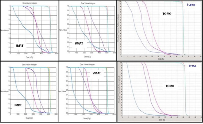
Figure 3. DVHs for the two different simulated positions in a typical case with plans by the three different techniques.

Blue line: small bowel; Pink line: femoral head; Purple line: bladder; Light blue line: PTV; Orange line: CTV.

Blue line: small bowel; Pink line: femoral head; Purple line: bladder; Light blue line: PTV; Orange line: CTV.