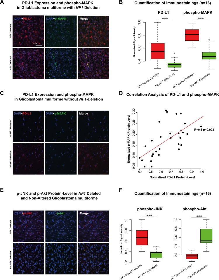
Figure 4.
A-C. Immunostaining of PD-L1 and phospho-MAPK14 in two patients with NF1 deletion (A) and two patient without NF1 alteration (C). Additionally, six independent fields were quantified by mean signal intensity by ImageJ, normalized and illustrated in a boxplot. D. Correlation analysis of protein-level of PD-L1 and p-MAPK showed a strong positive correlation (r=0.6 p<0.01). E-F. Immunostaining of phospho-JNK and phospho-AKT in a patient with NF1 deletion (upper panel) and one patient without NF1 alteration (lower panel). Additionally, quantification was given in the right boxplot (F). *** p<0.001, ** p=0.01, *p<0.05.

