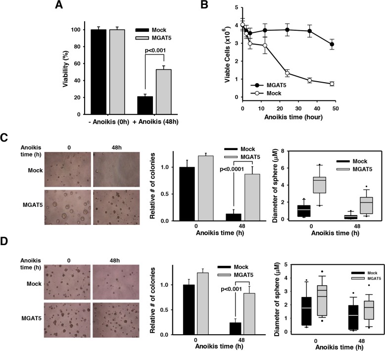
Figure 2. Anchorage-dependent and independent growth advantages by MGAT5-induced anoikis resistance.
A. Cell viability was assessed by an XTT assay to compare the anoikis resistance of mock and WiDr:MGAT5. Cells (1×10⁴) were placed into each well of 96-well plates and incubated in the presence of XTT solution for 2 hours. Absorbance at 450nm was measured in an ELISA reader (n=3). B. Cells were treated with anoikis stress for indicated times and allowed to form colonies on a culture plate. The number of colony was converted into viable cells by considering diluting factors (×1,000) (n=5). C-D. Cells were treated with anoikis stress for 48 hours and then embedded into soft agar (C) and basement membrane-based Matrigel (D). Cells were allowed to grow and the number of colonies and the diameter of colony spheres were measured (n=3).

