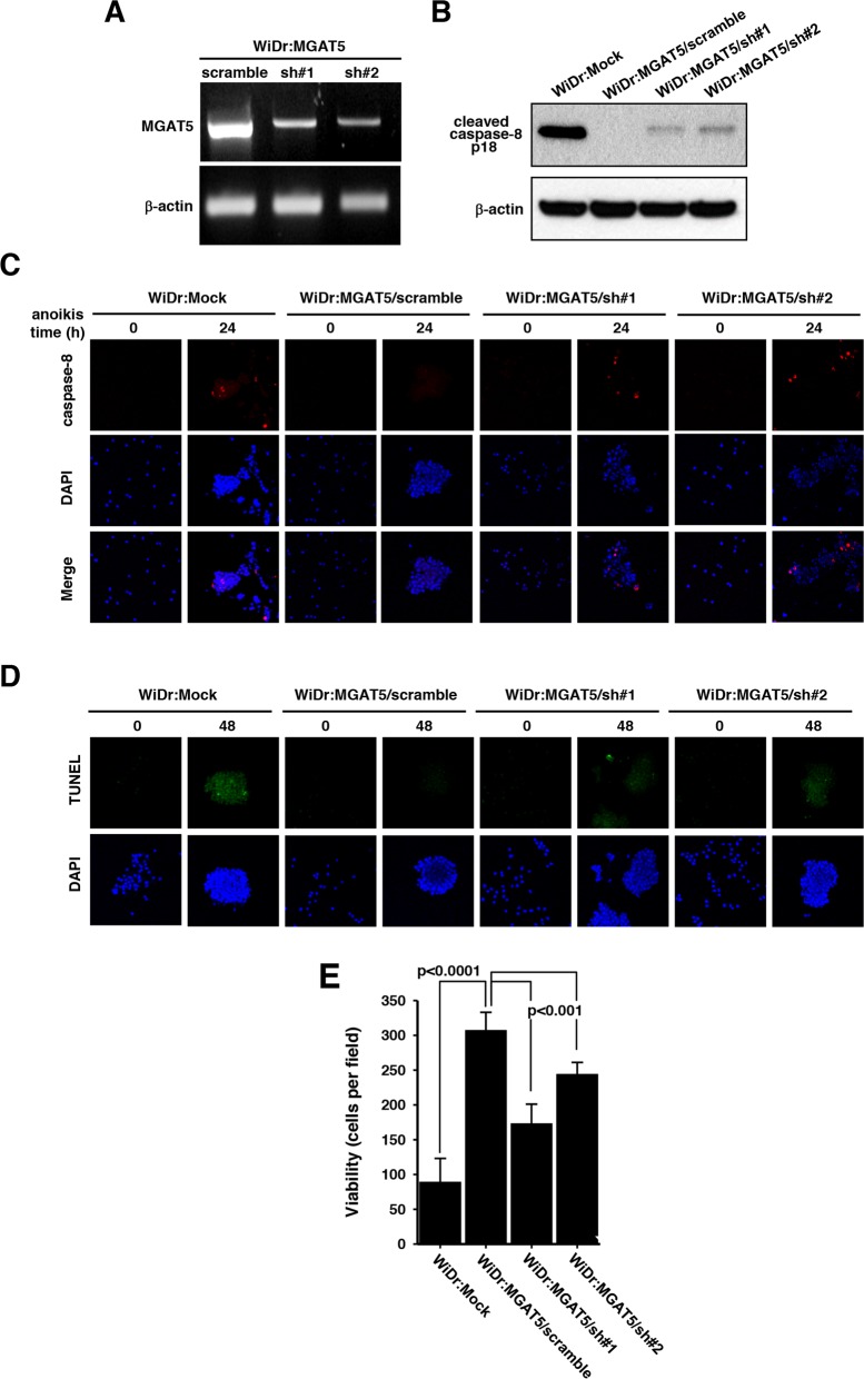
Figure 3. Sensitization of anoikis by suppressed MGAT5 expression.
A. The MGAT5 expression was suppressed through a small hairpin RNAs. The mRNA levels for two shRNA colonies were checked by RT-PCR followed on an agarose gel. B. Caspase-8 activation was compared among the mock, scramble, and MGAT5-suppressed cells by monitoring p18 products on an immunoblot. C-D. The apoptotic molecular signatures were monitored by caspase-8 activation through immunofluorescence (C) and by DNA fragmentation through TUNEL assay (D) following anoikis stress for 24-48 hours. E. Viability tests were performed by counting colonies formed on culture plates after anoikis stress for 48 hours. The values were averages of the number of colonies at 10 microscopic fields randomly selected at magnification of 400× (n=3).

