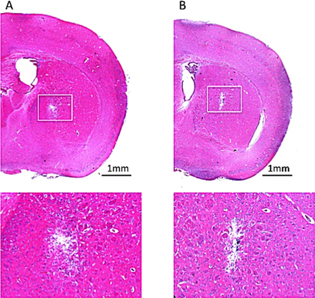
Figure 3. HE staining for histologically identifying potential tissue damage from the CED infused microbubbles.

(A) SonoVue with infused concentration of 0.5 mg/mL. (B) In-house fabricated Dox-MB with infusion concentration of 0.5 mg/mL. Bar = 1 mm.

(A) SonoVue with infused concentration of 0.5 mg/mL. (B) In-house fabricated Dox-MB with infusion concentration of 0.5 mg/mL. Bar = 1 mm.