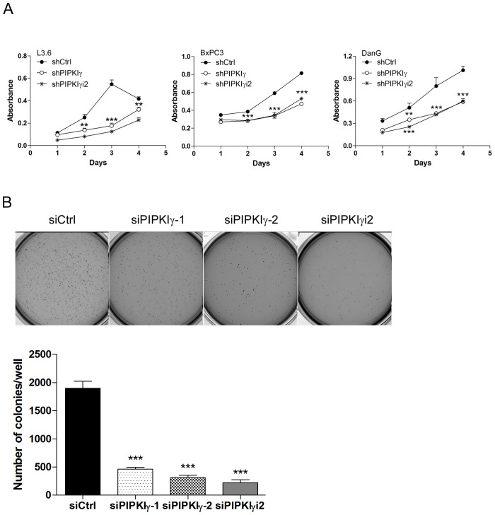
Figure 3. Depletion of PIPKIγ suppressed the proliferation and survival of PDAC cells.
Three different types of PDAC cells were infected with lentivirus carrying shRNA sequence for control (shCtrl), all PIPKIγ isoforms (sh IPKIγ), or PIPKIγi2 (shPIPKIγi2) for 48 hrs. Then cells were subjected to MTT assays at indicated time points (A). (B) L3.6 cells were transfected with control, PIPKIγ, or PIPKIγi2 siRNA; After 48 hours, soft agar colony formation was analyzed. Indicated L3.6 cells were harvested and suspended in culture medium containing 0.3% agarose, then plated in 6-well plates coated with 0.6% agar in triplicate. 14 days later, cells were stained with MTT and representative images from each group were taken and shown. Colony number in each well was quantified using GelCount software. Results from three independent experiments were statistically analyzed and plotted. ***, p<0.001.

