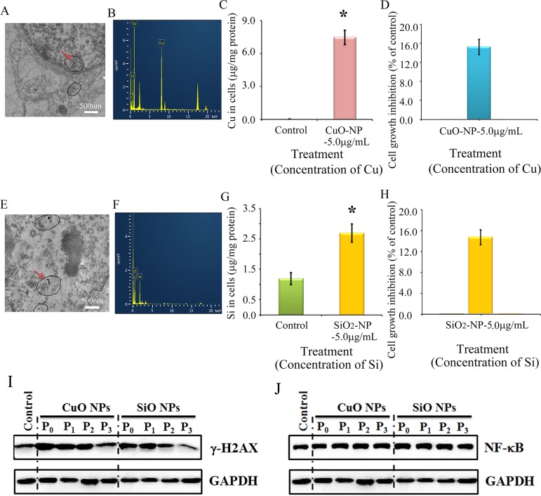
Figure 8. Impairment of γ-H2AX not NF-κB by CuO or SiO2 NPs in CHO-K1 cells.
(A) TEM photo of CuO NPs in CHO-K1 cells indicated by the red arrow; (B) EDS image of CuO NPs in CHO-K1 cells, two Cu peaks have shown; (C) Concentration of Cu in CuO NPs treated CHO-K1, *p < 0.05; (D) Effects of CuO NPs on CHO-K1 cell growth; (E) TEM photo of SiO2 NPs in CHO-K1 cells indicated by the red arrow; (F) EDS image of SiO2 NPs in CHO-K1 cells; (G) Concentration of Si in SiO2 NPs treated CHO-K1, *p < 0.05; (H) Effects of SiO2 NPs on CHO-K1 cell growth; (I) Decrease in γ-H2AX by CuO and SiO2 NPs using WB analysis; (J) No alteration in NF-κB major component p65 by CuO NPs or SiO2 NPs treatment using WB analysis; N ≥ 3.

