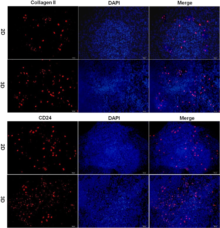
Figure 6. Immunofluorescence analysis of iPSC differentiation in hydrogel.
Immunofluorescence staining of iPSCs for the presence of collagen II and CD24. Positive staining is shown by red fluorescence, blue fluorescence is for the nucleus (magnification 200×, bar = 50 μm). 2D: iPSCs differentiated in culture plate, 3D: iPSCs differentiated in hydrogel.

