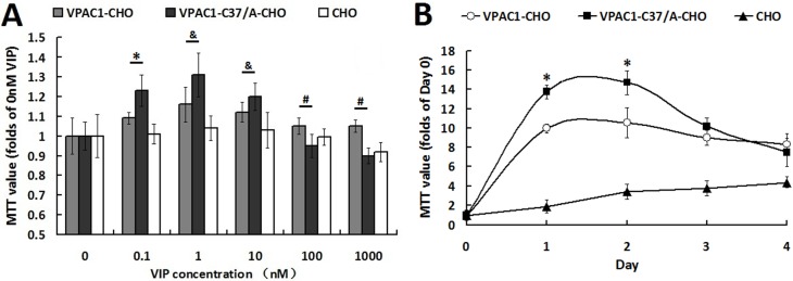
Figure 2. VPAC1-CHO cells had lower proliferative activity than VPAC1-C37/A-CHO cells.
(A) The cell viabilities assayed by MTT methods of VPAC1-CHO, VPAC1-C37/A-CHO and CHO cells incubated with VIP (0.1 nM–1000 nM). VPAC1-C37/A-CHO cells proliferated more rapidly than VPAC1-CHO cells when incubated with low concentration of VIP (0.1 nM–10 nM) (*p < 0.01, VPAC1-C37/A-CHO vs. VPAC1-CHO; &p < 0.05, VPAC1-C37/A-CHO vs. VPAC1-CHO), but VPAC1-CHO cells remained higher viability than VPAC1-C37/A-CHO cells when incubated with high concentration of VIP (100 nM–1000 nM) (#p < 0.01, VPAC1-CHO vs. VPAC1-C37/A-CHO). The data were means ± SEM of six experiments. (B) The growth curve of VPAC1-CHO, VPAC1-C37/A-CHO and CHO cells with 0.1 nM VIP for 4 days. VPAC1-C37/A-CHO cells proliferated more rapidly than VPAC1-CHO cells before the logarithmic phase (*p < 0.01, VPAC1-C37/A-CHO vs. VPAC1-CHO), but faded more rapidly than VPAC1-CHO cells after the logarithmic phase. The data were means ± SEM of six experiments.

