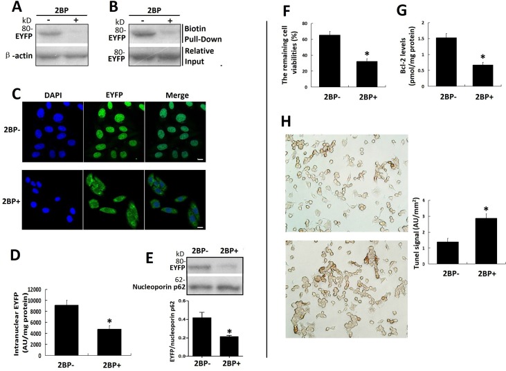
Figure 7. Palmitoylation inhibitor 2-BP inhibited the nuclear translocation of VPAC1-EYFP and the anti-apoptotic activity of VPAC1-CHO cells synchronously.
(A) The acyl-biotin exchange assay was used to confirm the inhibitory effects of 2-BP on the palmitoylation of VPAC1-EYFP. (B) The click chemistry palmitolaytion assay was used to confirm the inhibitory effects of 2-BP on the palmitoylation of VPAC1-EYFP. (C) Confocal fluorescence micrographs showed the fluorescence signal representing VPAC-EYFP internalized but did not further transfer into nucleus while incubated with 2-BP, indicating that 2-BP inhibited the nucleus translocation of VPAC1-EYFP induced by VIP. Bar, 5 μm. (D) Intranuclear fluorescence quantification showed the intranuclear fluorescence signal of VPAC1-EYFP in treatment with 2-BP (2BP+) was significantly lower that that in treatment with without 2-BP (2BP-) (*P < 0.01, 2BP+ vs. 2BP-). The data were means ± SEM of six experiments. (E) Western blotting of nucleoprotein showed that the amount of the intranuclear VPAC1-EYFP was significantly decreased by treatment with 2-BP (*P < 0.01, 2BP+ vs. 2BP-). The data were means ± SEM of six experiments. (F) The treatment with 2-BP decreased the percentage of the cell remaining viability of VPAC1-CHO cells (*P < 0.01, 2BP+ vs. 2BP-). The data were means ± SEM of six experiments. (G) The treatment with 2-BP decreased the intracellular anti-apoptotic protein Bcl-2 level of VPAC1-CHO cells. (*P < 0.01, 2BP+ vs. 2BP-). The data were means ± SEM of six experiments. (H) Microscope imaging of VPAC1-CHO cells treated with 2-BP (2BP+) or without 2-BP (2BP-) were dyed by TUNEL assay. It was shown that brown staining in VPAC1-CHO cells treated with 2-BP was significantly more than that treated without 2-BP, which was confirmed by the statistic analysis on the TUNEL signals (*P < 0.01, 2BP+ vs. 2BP-). The data were means ± SEM of six experiments.

