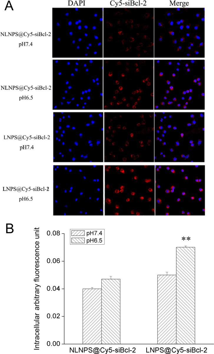
Figure 5. The cellular uptake of LNPS@Cy5-siBcl-2 and NLNPS@Cy5-siBcl-2 on A549 cells in 4 h.
(Panel A) are the cellular uptake of LNPS@Cy5-siBcl-2 and NLNPS@Cy5-siBcl-2 detected by CLMS in pH7.4 and pH6.5 culture medium. (Panel B) is the statistic result of panel A. 20 × oil immersion objective and 10 × ocular lens. Red indicates Cy5-siBcl-2, and blue indicates nucleus. Data are expressed as the mean ± SD, n = 3. **p < 0.01, vs NLNPS@Cy5-siBcl-2 in the same pH value.

