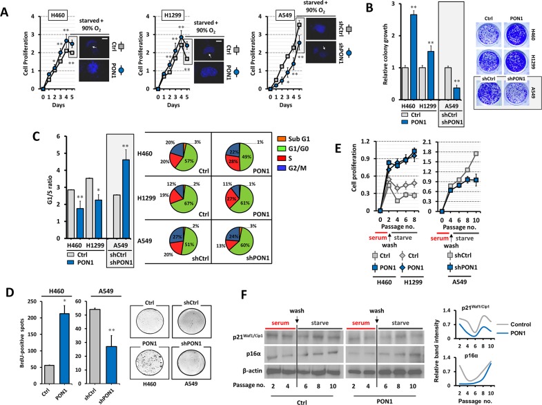
Figure 3. PON1 regulation impacts lung cancer cell growth and arrest programs.
(A) Assessment of cell viability. Before day 5, cells were serum starved for 24 h and were subjected to DAPI staining. Subsequent to serum starvation, cells were pre-exposed to 90% O2 level for 12 h and incubated in normoxia for 12 h (displayed as immunofluorescence in right panel of each graph). (B) Colony formation (>50 cells) was assessed by crystal violet staining. (C) Mean G1/S-phase ratio from cell cycle analysis (left panel) and cell cycle progression fractions of >15,000 cells analyzed by FACS (right panel). (D) BrdU-positive spots of cells examined at day 4 at early cell passage in serum-starved medium. (E) Cell passage-dependent viability after pre-culture of cells at low density for 3 days in serum-rich media containing low etoposide concentration and then subsequently starved them for another 6 days. SA-β-gal assay was used to confirm presence of senescent cells after recovery (data not shown). (F) Western blot analysis of whole cell lysates of same samples from E. β-actin served as the loading control. *P<0.05. Error bars are mean ± S.E.M. n=3~5. All experiments were conducted at least in triplicate and biologically repeated at least twice. Statistical analysis was performed using unpaired Student's t test.

