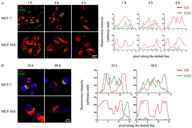
Figure 2. WVLGE-containing polypeptide enters into breast cancer cells.

(A) and (B) MCF-7 and MCF-10A cells were incubated with FITC-labeled WVLGE-containing azurin CPP for 24 hours and stained by membrane dye DiI for 15 minutes. Cells were incubated in fresh medium for different times as indicated before observation by confocal microscopy. Dapi was used to stain the nuclear for observation at 24 hours and 48 hours (B). Scale bars are 20 μm (A) and 10 μm (B), respectively. The fluorescence intensities of each pixel along the white dotted line are plotted. The X-axis represents the pixels along the white dotted line and the Y-axis represents the fluorescence intensity.
