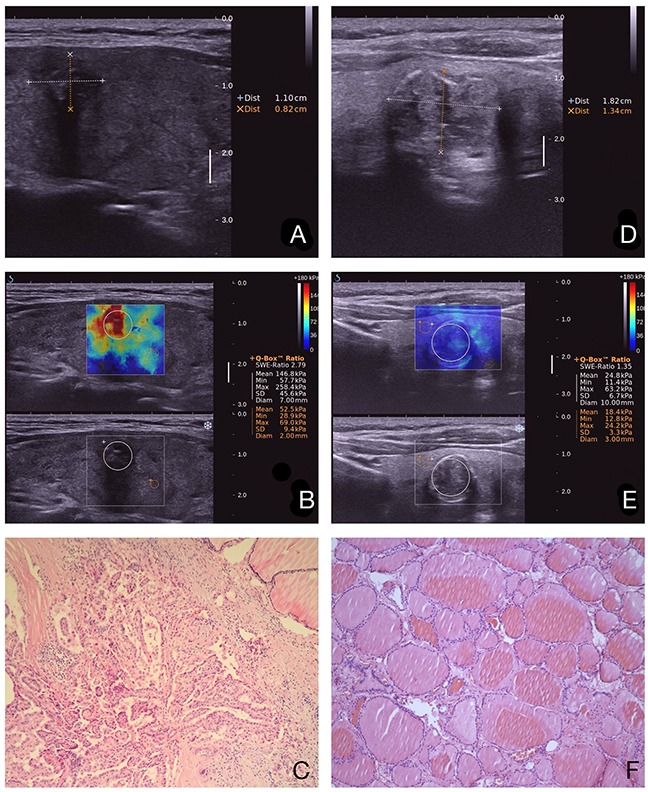
Figure 3. Images in a 64-year-old woman who underwent a routine checkup.

An 11-mm left thyroid solid nodule with marked hypoechogenicity, poorly defined margins, and micro-calcifications was found on gray-scale US, was classified as TI-RADS 4c a. The Emax value of SWE of the nodule was 258.4 kPa b. This thyroid nodule was diagnosed as papillary thyroid carcinoma after surgery. Pathological images (c. HE 10×10). Images in a 59-year-old woman who underwent a routine checkup. An 18-mm left thyroid nodule with macro-calcification was found on conventional US, was classified as TI-RADS 4a d. The Emax value of SWE of the nodule was 63.2 kPa e. The post-operational histopathology was nodule gotiers. Pathological images (f. HE 10×10).
