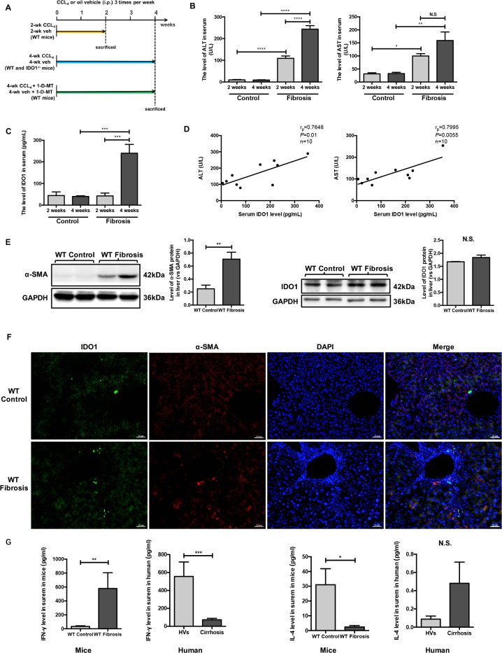
Figure 2. Serum IDO1 was time-dependently elevated in a CCl4-induced fibrosis mouse model and positively correlated with liver lesions.
(A) Experimental plan for mice. (B) Liver lesions were assessed by measuring serum ALT and AST levels in WT control and model mice. (C) ELISA for detecting serum IDO1 level in WT control and model mice. (D) Pearson's linear correlation tests for serum IDO1, ALT and AST levels in the WT model mice. (E) Western blot analysis of the expression of IDO1 and α-SMA in the livers of WT control and model mice (4-week). (F) Immunofluorescence analysis to detect the expression of IDO1 and α-SMA in the livers of WT control and model mice (4-week). (G) ELISA for detecting serum IFN –γ and IL-4 in mice and clinical subjects. The data are presented as the means ± SEM (*P < 0.05, **P < 0.01, ***P < 0.001, ****P < 0.0001).

