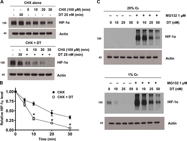
Figure 3. DT inhibits hypoxic HIF-1α accumulation by inhibiting protein synthesis.

(A) GSC were treated with solvent alone (CHX; top panel) or 25 nM DT (CHX + DT; bottom panel) for 1 h, followed by incubation with 100 μM CHX from 0 to 30 min. Cell lysates were subjected to immunoblotting using antibodies against HIF-1α or actin. (B) Intensity of HIF-1α protein signals obtained in A was quantified using Eagle Sight densitometry software (Version 3.21; Stratagene). The HIF-1α densitometry data were normalized to those of the control (Lane 1) and actin levels. The plots represent the mean ± SD from three independent experiments. Calculation of HIF-1α half-life was performed by the regression program of Microsoft Excel 2000. (C) Cells were treated with 10–50 nM DT in the presence or absence of 1 mM MG132 in 20% and 1% O2 for 8 h. Cell lysates containing equal amounts of protein (20 mg) were separated by SDS-PAGE and immunoblotted with an anti-HIF-1α antibody. Actin was used as a loading control.
