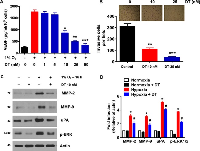
Figure 4. DT inhibits GSC angiogenic and invasive capacities.

(A) GSC were treated with various concentrations of DT (1–50 nM) for 16 h during hypoxia. Concentration of VEGF protein in the culture medium was determined by ELISA. The assays were performed in triplicates. (B) GSC were treated with DMSO or DT, and were cultured in sphere forming conditions in the upper well of matrigel-precoated transwell chambers for 16 h. Cells invading the lower well were fixed and stained with a Diff-Quick kit and photographed (100× magnification). Invasiveness was determined by counting cells in five randomly selected microscopic fields per well. (C and D) Western blot analysis shows that DT downregulates expression of invasion-related proteins in GSC during hypoxic conditions. Error Bars represent mean ± SD of triplicate samples. *P < 0.05, **P < 0.01, and ***P < 0.005.
