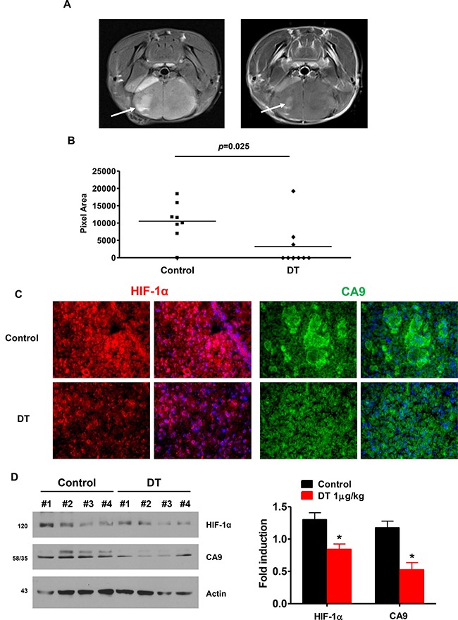
Figure 7. DT treated GSC have reduced capacity for tumor formation and attenuates HIF signaling in an orthotopic model.

(A) Tumor formation was monitored by MRI Arrow points to tumor. (B) Quantification of MR imaged tumor size. DT significantly reduced tumor size (p = 0.025). (C) HIF-1α and CA9 immunostaining in brain sections from vehicle- and DT-treated mice. (D) Anti-HIF-1α and anti-CA9 expression was assessed in four different protein samples using western-blotting (right), and the average value is shown on left. All experiments with statistical analyses were performed at least three times, and error bars depict means ± SD; *P < 0.05.
