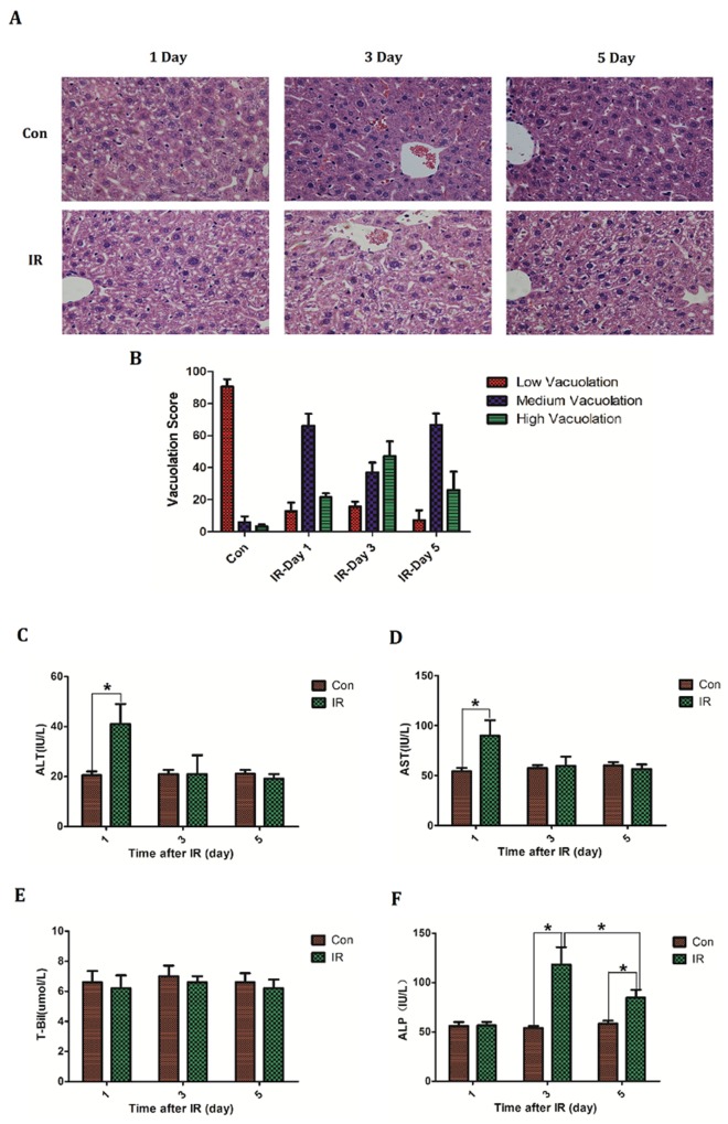
Figure 1. The establishment of acute radiation-induced liver injury model of mice.
BABL/C mice received hepatic 30Gy of irradiation. On the first, third and fifth day after irradiation the livers were taken to (A, B) make histologic evaluation by H&E staining (n=5) (micrographs in A; quantified in B); Bloods were collected to measure (C) ALT level, (D) AST level, (E) T-Bil level and (F) ALP level (n=5). Magnification × 200. Data were presented as mean± SD. *P<0.05.

