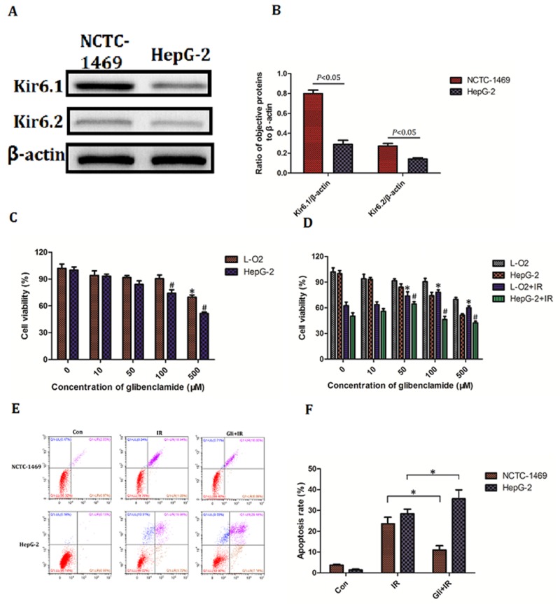
Figure 7. Effect of glibenclamide on hepatoma carcinoma cell.
(A) Human hepatic cell line L-O2 and human hepatoma cell line HepG-2 was harvested to detect the protein expression of Kir6.1 and Kir6.2 by western blotting. (B) Quantitative analysis were performed (n=3). (C, D) L-O2 and HepG-2 were treated with different concentration of glibenclamide for 1 hour. 24 hour after combining with/without IR the cells viability was determined by CCK-8 assay. (E, F) L-O2 and HepG-2 were pre-treated with 100μM glibenclamide for 1 hour. 48 hour after 8Gy of irradiation the cells apoptosis rate were tested by flow cytometry (n=3). Data were presented as mean± SD. *P<0.05 VS L-O2 at 0μM glibenclamide; #P<0.05 VS HepG-2 at 0μM glibenclamide.

